

To look at the difference between main and secondary retinal ganglion cell (RGC) degeneration, the protein expression at 4 areas of retina together with superior, temporal, inferior and nasal quadrant in a rat mannequin of partial optic nerve transection (pONT) using 2‑D Fluorescence Difference Gel Electrophoresis (DIGE) had been investigated.
Unilateral pONT was carried out on the temporal aspect of optic nerves of grownup Wistar rats to separate main and secondary RGC loss.
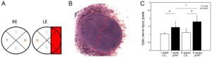
Topographical quantification of RGCs labeled by Rbpms antibody and evaluation of axonal harm by grading of optic nerve harm at 1 week (n=8) and eight weeks (n=15) after pONT demonstrated early RGC loss at temporal area, which is taken into account as main RGC degeneration and progressing RGC loss at nasal area, which is taken into account as secondary RGC degeneration.
Early protein expression in every retinal quadrant (n=4) at 2 weeks after pONT was in contrast with the corresponding quadrant in the contralateral management eye by DIGE. For all comparisons, 24 differentially expressed proteins (>1.2‑fold; P<0.05; ≥three non‑duplicated peptide matches) had been recognized by mass spectrometry (MS).
Interestingly, in the nasal retina, serum albumin and members of crystallin household, together with αA, αB, βA2, βA3, βB2 and gamma S indicating stress response had been upregulated. By distinction, solely αB and βA2 crystallin proteins had been altered in temporal quadrant.
In the superior and inferior quadrants, βB2 crystallin, keratin kind I, S‑arrestin and lamin‑B1 had been upregulated, whereas warmth shock cognate 71 kDa protein and heterogeneous nuclear ribonucleoproteins A2/B1 had been downregulated. In abstract, the use of DIGE adopted by MS is beneficial to detect early regional protein regulation in the retina after localized optic nerve harm.
A Quantitative Proteomics Study of Early Heat-Regulated Proteins by Two-Dimensional Difference Gel Electrophoresis Identified OsUBP21 as a Negative Regulator of Heat Stress Responses in Rice.
To perceive the early warmth shock (HS)-regulated mobile responses that affect the tolerance of rice plant to excessive environmental temperatures, two-dimensional difference gel electrophoresis (2D-DIGE) is carried out to discover the early HS-regulated proteome.
Multiple proteins that present abundance modifications after 1 and 5 min of HS remedy are recognized. Of the early HS-regulated proteins recognized, the abundance of a ubiquitin-specific protease, OsUBP21, and its Arabidopsis homolog, AtUBP13, is discovered to be upregulated by 5 min of HS remedy.
Further, knocking the expression of OsUBP21 or AtUBP13 down or out will increase the tolerance of rice and Arabidopsis crops to HS stress, suggesting that the operate of these ubiquitin-specific proteases in regulating plant HS responses is conserved between monocots and dicots.
2D-DIGE confirmed a gaggle of proteins are differentially regulated in wild-type and ubp21 mutant after 30 min of HS remedy.
Among these proteins, 11 are discovered to work together straight with OsUBP21; thus, they could be targets of OsUBP21. Future analyses of the roles of these OsUBP21-interacting proteins in plant HS responses will assist reveal the protein ubiquitination/deubiquitination-regulated mobile responses induced by HS in rice.